The future of memory: Neuroimaging memory and imagination with functional MRI
Professor Donna Rose Addis, School of Psychology, Faculty of Science; Associate Director, Centre for Brain Research, University of Auckland
Overview
Neuroimaging is a critical method for understanding brain function and the field of cognitive neuroscience. In the Memory Lab, we use functional magnetic resonance imaging (fMRI) to measure the blood oxygenation level dependent response (the fMRI signal) to cognitive tasks such as remembering the past and imagining the future. This work has been at the forefront of a reconceptualisation of memory to now consider how memory contributes to many aspects of cognitive functioning and psychological wellbeing, including how we simulate future events, construct a sense of identity and how we generate creative ideas. Moreover, fMRI enables us to investigate how these abilities change in healthy ageing, dementia and depression.
Remembering the past
Crucial remembering the past in rich detail is the hippocampus: more vividly detailed memories are associated with higher levels of hippocampal activity and hippocampal damage results in reduced episodic detail. However, the hippocampus doesn’t work in isolation, and is part of a whole-brain network supporting AM retrieval. We have found that distinct parts of this brain network are associated with different types of memory (e.g., specific vs. general AMs; Addis et al., 2011) and different “routes” to recovering memories (e.g., generative vs. direct retrieval; Addis et al., 2012).
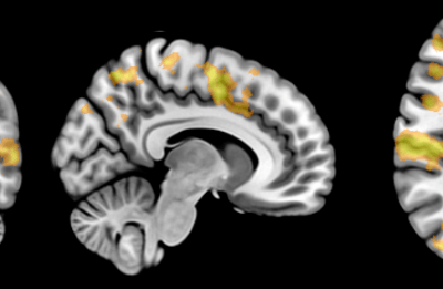
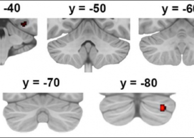
Currently, we are exploring the contributions of other parts of this network to AM retrieval, such as the cerebellum. In a recent Activation Likelihood Estimation (ALE) meta-analysis, we show that the right posterior cerebellum is reliably activated in studies of remembering, and this region is a node of the autobiographical memory retrieval network (Figure 1, Addis et al., 2016).
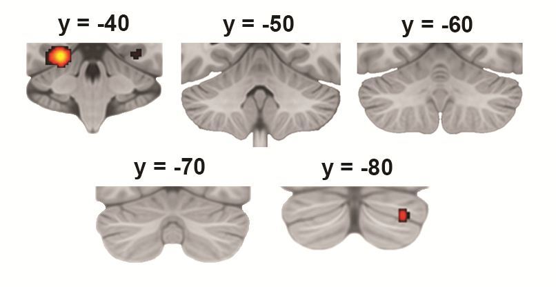
Figure 1. Cerebellar activity while remembering past events as revealed by Activation Likelihood Estimation (ALE) meta-analysis (Addis et al., 2016, Neuropsychologia).
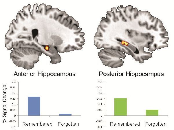
Figure 2. Anterior and posterior hippocampal activity during the successful encoding of future simulations that are later remembered, as compared to events that are later forgotten
(Martin et al., 2011, PNAS).
Imagining the future
Our work on this topic started with the striking finding that the brain activity during future simulation was incredibly similar to that during remembering, indicating shared neural and cognitive processes. In addition to findings of striking overlap of the neural correlates of remembering and imagining, our neuroimaging research has consistently found increased hippocampal activity, suggest that future simulation is a more intensive process than remembering. In a number of fMRI studies conducted at the Memory Lab, we have demonstrates that this preferential hippocampal activity during simulation is related to the novelty of imagined events (van Mulukom et al., 2013) as well as the successful encoding of these events into memory (Figure 2, Martin et al., 2011). Recently, we have begun to explore other factors that contribute to imagination, including creativity.
Neuroimaging analysis methods
The Memory Lab is also interested in neuroimaging analysis methods. In a recent study, we demonstrated that task-related functional connectivity is dependent on whether the analysis is conducted within-subjects or across subjects; we also introduced a novel measure within-subjects task-related functional connectivity (Roberts et al., 2016). We are currently exploring a novel functional MRI measure of cognitive capacity – BOLD variability – and how this is related to measures of blood flow.
Resources & facilities at the Centre for eResearch
The Centre for eResearch has worked with our research group over the past five years to achieve a number of outcomes for both our research projects and our research-led teaching. Initially CeR transferred data and to a newly created virtual machine with fMRI software requiring graphical multistep analysis with access by multiple users (from different locations around the users). They also assisted us in testing the computational requirements of our analyses to design the most appropriate solutions. They created a system where it was easy to copy data from other locations such as the CAMRI instrument servers. The VM and the data is backed up externally on other network storage with automatic syncing overnight.
Designing an archival system and computational infrastructure
More recently we have been working with CeR to design a system for a centralized repository of University MRI data collected by researchers across the Centre for Brain Research (including academics in FoS and FMHS). The infrastructure will involve a series of VMs for users as well as integrated data storage in the form of an XNAT archive – a searchable archive of raw and derived MRI data that facilitate easy data sharing with researchers across UoA as well as other institutions (Figure 3). This project has involved consultation and design work by CeR with central teams on the specifics of server specifications and options.
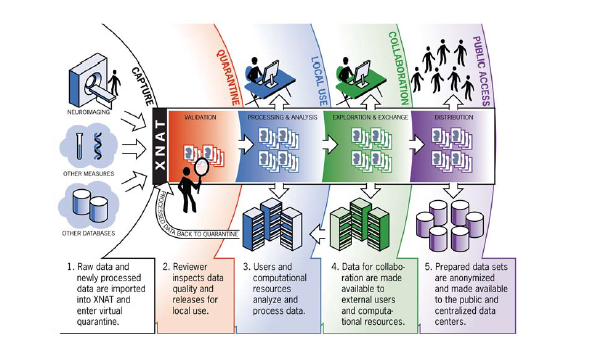
Figure 3. XNAT will serve as a hub for capturing MRI and related data from multiple sources and distributing it to a variety of end users (Marcus et al., 2011, Neuroinformatics).
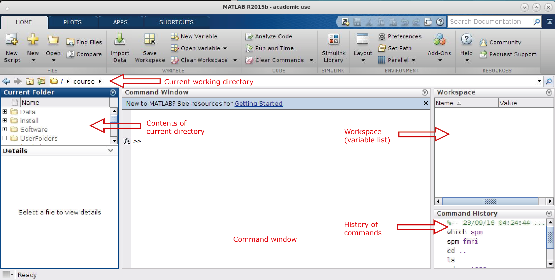
Figure 4. Screenshots provided to students in the PSYCH727 Lab Manual Matlab.
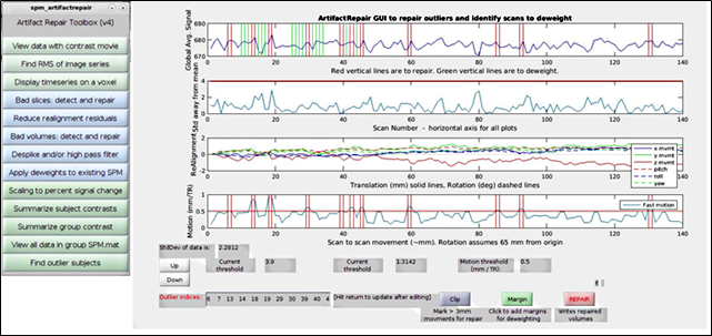
Figure 5. Screenshots provided to students in the PSYCH727 Lab Manual: ArtRepair.
Teaching fMRI to graduate classes
Additionally, graduate students often come to the lab lacking the in-depth overview, digital and statistical skills required to understand and analyse fMRI data. This year, a 700 level course was introduced in Psychology (PSYCH727) with a particular focus on fMRI use in cognitive neuroscience. This course, designed for beginners, provided comprehensive coverage of experimental design, image acquisition, image pre-processing, analysis methods (ranging from univariate GLM approaches to multivariate and connectivity approaches), localisation and interpretation.
Classes involved a lecture followed by a hands-on laboratory working with fMRI data (with assistance from instructors) to consolidate learning of concepts and techniques. Staff at the Centre for eResearch architected, provisioned, and installed software and storage mechanisms on research virtual machines to make it possible for this research-led teaching course to provide the students with a true research experience. Students worked with a class-sized data sets, with local storage allocated in personal folders. Graphical interfaces were provided using an x2GO virtual desktop experience with connections form clients in lab environments. Students were introduced to Linux and Matlab, and provided with basic knowledge of relevant commands. Analysis was undertaken using toolsets such as Matlab, spm12, ArtRepair, MRIcron, PLS and REX (Figures 4 and 5).
PSYCH727 has been a resounding success and has provided Psychology students with a strong foundation for the use of fMRI data (as well as Linux and Matlab) in cognitive neuroscience research. These skills will support their postgraduate research at Honours, Masters and Doctoral levels.
See more case study projects

Our Voices: using innovative techniques to collect, analyse and amplify the lived experiences of young people in Aotearoa
Painting the brain: multiplexed tissue labelling of human brain tissue to facilitate discoveries in neuroanatomy

Detecting anomalous matches in professional sports: a novel approach using advanced anomaly detection techniques
Benefits of linking routine medical records to the GUiNZ longitudinal birth cohort: Childhood injury predictors
Using a virtual machine-based machine learning algorithm to obtain comprehensive behavioural information in an in vivo Alzheimer’s disease model
Mapping livability: the “15-minute city” concept for car-dependent districts in Auckland, New Zealand
Travelling Heads – Measuring Reproducibility and Repeatability of Magnetic Resonance Imaging in Dementia
Novel Subject-Specific Method of Visualising Group Differences from Multiple DTI Metrics without Averaging

Re-assess urban spaces under COVID-19 impact: sensing Auckland social ‘hotspots’ with mobile location data
Aotearoa New Zealand’s changing coastline – Resilience to Nature’s Challenges (National Science Challenge)
Proteins under a computational microscope: designing in-silico strategies to understand and develop molecular functionalities in Life Sciences and Engineering

Coastal image classification and nalysis based on convolutional neural betworks and pattern recognition
Determinants of translation efficiency in the evolutionarily-divergent protist Trichomonas vaginalis
Measuring impact of entrepreneurship activities on students’ mindset, capabilities and entrepreneurial intentions

Using Zebra Finch data and deep learning classification to identify individual bird calls from audio recordings
Automated measurement of intracranial cerebrospinal fluid volume and outcome after endovascular thrombectomy for ischemic stroke
Using simple models to explore complex dynamics: A case study of macomona liliana (wedge-shell) and nutrient variations
Fully coupled thermo-hydro-mechanical modelling of permeability enhancement by the finite element method
Modelling dual reflux pressure swing adsorption (DR-PSA) units for gas separation in natural gas processing
Molecular phylogenetics uses genetic data to reconstruct the evolutionary history of individuals, populations or species
Wandering around the molecular landscape: embracing virtual reality as a research showcasing outreach and teaching tool
