Using research virtual machines to analyse fMRI datasets
Dr Reece Roberts, Research Fellow, Psychology
Introduction
Magnetic resonance imaging (MRI) allows clinicians and researchers to leverage differences in the magnetic properties of different tissue types and/or blood to generate images of brain structure and function (fMRI). A number of research groups at The University of Auckland, in both the Faculties of Science and Medical and Health Sciences collect MRI data of various types at the Centre for Advanced Magnetic Resonance Imaging (CAMRI; https://www.fmhs.auckland.ac.nz/en/faculty/camri.html). In the case of fMRI, these data are 4D datasets comprised of a series of 3D images (each acquired every 1-3 seconds). A number of research groups have collaborated with CeR to build virtual machines suited to analysing these large, complex datasets.
Data organisation
An increasingly common way to structure data is to use the Brain Imaging Data Structure format (BIDS; http://bids.neuroimaging.io/), which is a movement to standardise data structures across the worldwide neuroimaging community, to allow for greater sharing of data and an increase in reproducibility of neuroimaging research.
Data quality assurance and preprocessing
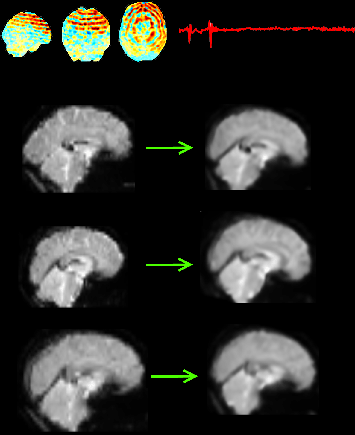
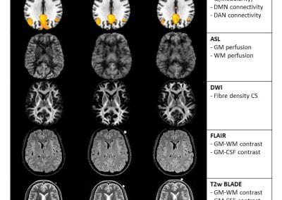
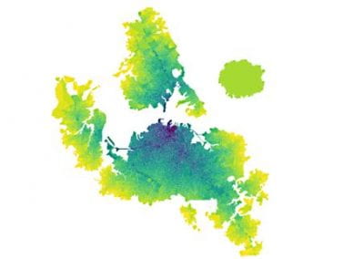
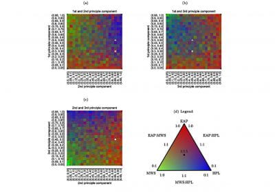
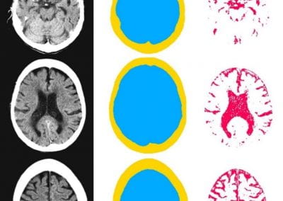
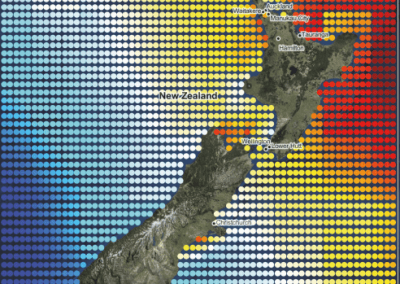
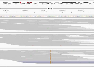
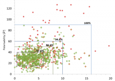
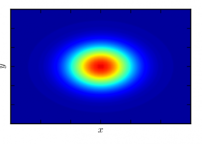
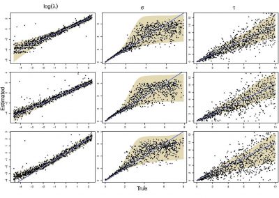
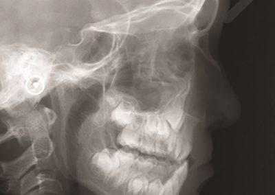
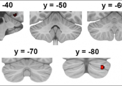
Adopting the BIDS data structure allow for the use of a number of open-source packages to be applied to a given dataset. For example, automated quality assurance of MRI images (https://github.com/poldracklab/mriqc) produces metrics enabling the detection of outliers on a range of measures (see Fig. 1).
To enable the analysis of group effects, MRI scans from study participants are preprocessed (using, e.g., fMRIprep; https://github.com/poldracklab/fmriprep) to remove sources of variance that are unrelated to the signal of interest (e.g. motion and scanner artefacts, physiological noise). In addition, the images of each individual are normalised to be in a common space. Performing these analyses on CeR VMs has required the installation of Docker packages, and CeR staff have been critical in getting these up-and-running. This is particularly helpful as Docker containers are not currently supported on NeSI HOC platform.
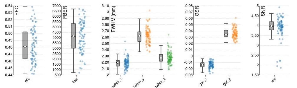
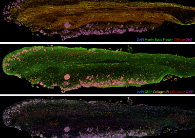
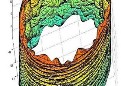
Fig. 1. Some example parameters from automatic quality assurance of MRI images that allow for automatic rejection of outliers.
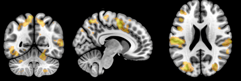
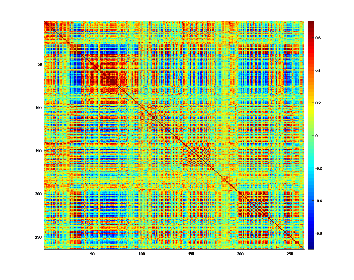
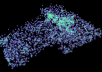
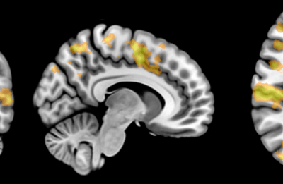
Fig. 3. On the top, a statistical parametric map showing widespread increases in brain activity in response to a cognitive task. At the bottom, a correlation matrix of 264 regions of interest (ROIs) showing the strength of the temporal correlation between the time-series